SPECIALIZED EQUIPMENT
The latest diagnostic equipment to provide the most efficient and accurate eye examination.Listowel Vision Care has the latest diagnostic equipment to provide the most efficient and accurate eye examination. Combining state-of-the-art technology with clinical experience allows us to diagnose and manage all of your eye care needs. Our advanced testing also allows us to have digital images stored to compare them with the results of future tests. This allows us to detect minute changes that we may not have been able to identify without these tests. We strongly recommend advanced testing to all of our patients.
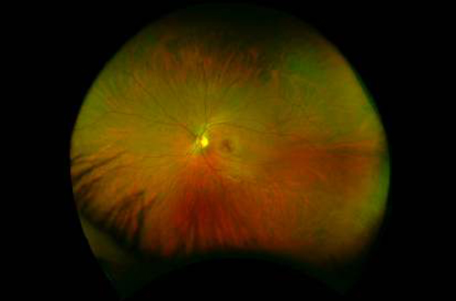
optomap Ultra-widefield retinal imaging

Healthy Eye

AMD
Optos introduced ultra-widefield (UWF™) retinal imaging to enable eyecare professionals to discover, diagnose, document and treat ocular pathology that may first present in the periphery – pathology which may go undetected using traditional examination techniques and equipment. The UWF, high-resolution retinal imaging devices each image more than 80% or 200˚ of the retina in a single capture. With optomap auto-montage, up to 97% or 220⁰ of the retina can be imaged with the multi-capture, montaging functionality.

The Benefits of optomap
optomap offers unparalleled views of the retina which provide eye care professionals with the following: The only anatomically correct 200⁰ or 82% image of the retina Simultaneous view of the central pole, mid-periphery and periphery Integrated hardware & software platform Multiple retinal imaging modalities to see more, discover more, and treat more patient diseases and pathologies, more effectively With more than 600 published and ongoing clinical trials, as well as thousands of case studies and testimonials, show the long-term value of optomap imaging in diagnosis, treatment planning, and patient engagement.
Unlike full spectrum white light used in conventional devices, optomap technology incorporates low-powered laser wavelengths that scan simultaneously. This allows review of the retinal substructures in their individual laser separations. 1
Digital Retinal Imaging

A picture is worth a thousand words. Now we can take a digital photograph of the important retinal structures that line the inside the eye, including your optic nerve and vascular branches. Through viewing the digital photo we can see whether the eye is healthy or if there are any abnormalities that may threaten vision or require attention and monitoring. It is especially recommended for patients with a history of diabetes, glaucoma, macular degeneration or any other retinal disorders.
These photographs provide a great baseline for comparison during future visits to help maintain a lifetime of healthy vision. The screening only takes a few minutes to complete and the digital image can be viewed immediately by both the doctor and the patient at the time of the visit, allowing for quick diagnosis. We believe that all of our patients should be screened with a digital photo, as it will help us provide the best eye care for you and your loved ones.
Optical Coherence Tomography (OCT SLO)

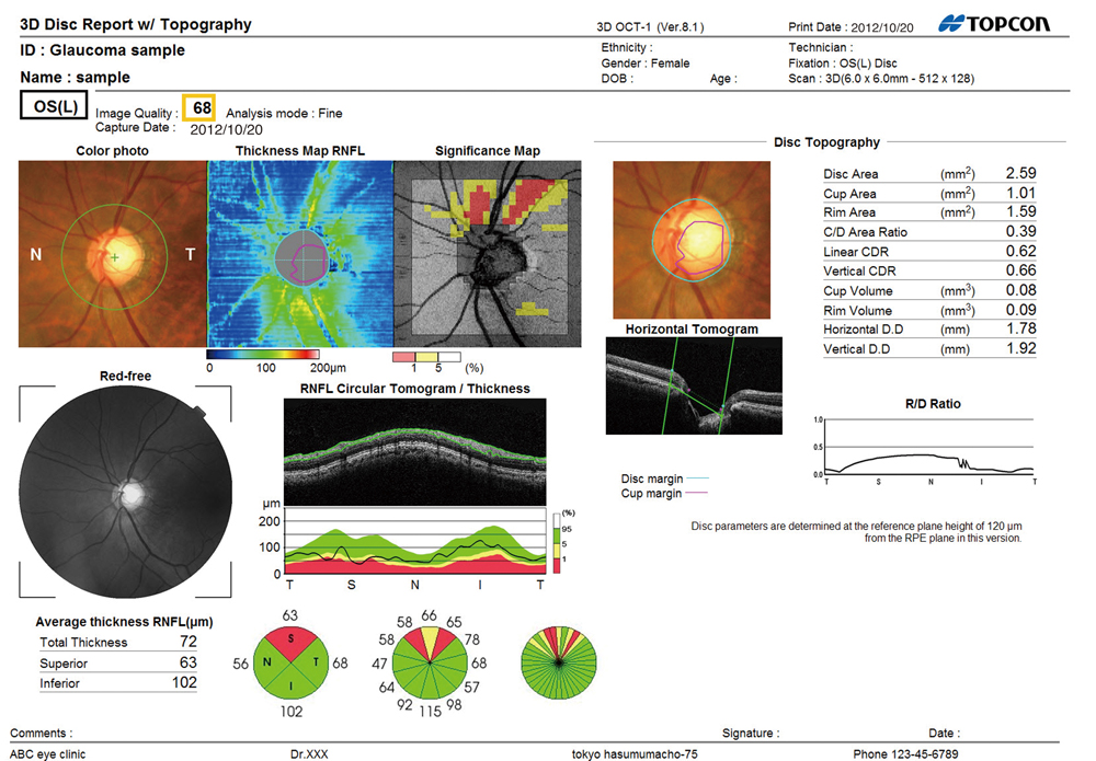
We offer the latest in ocular imaging with a 3D Optical Coherence Tomography (OCT SLO). This state-of-the-art diagnostic machine uses an advanced imaging technique allowing us to see the structures of your retina and optic nerve in cross-section, like an MRI. Since images can be taken easily, the OCT is useful in the detection of many ocular diseases and can be performed as a screening test. The acquisition of OCT scans is quick, completely non-invasive and painless. We can analyze each of the retina’s distinctive layers, allowing our doctors to accurately locate any abnormalities and measure your retinal thickness. This aids in the accurate diagnosis and management of glaucoma, macular degeneration, diabetic retinopathy, post-surgery edema, and other retinal diseases. If this test is recommended by your doctor or if you choose to add an OCT scan to your routine examination, the additional costs will be explained to you prior to any testing.
Topcon TRK-1P Autorefractor and Non-Contact Tonometry/Pachymetry
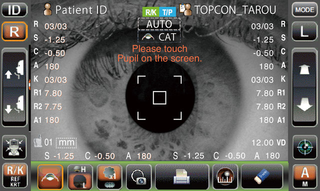

The Topcon TRK-1P is a fully automated non-contact system that quickly and accurately measures the surface of the eye and estimates a patient’s refractive error with one push of a button. It simultaneously measures the pressure of the eye and corneal thickness, which helps in the assessment and management of anterior surface conditions. It also helps our clinicians to fit contact lenses more effectively, monitor for ocular diseases and provide the most accurate spectacle prescriptions possible.
Visual Field Analyzer
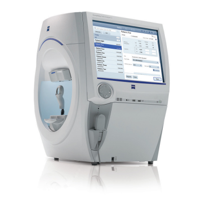
The Humphrey Visual Field Analyzer is the gold standard for detecting and monitoring glaucoma. This tool allows our clinicians to quickly and accurately analyze your field of vision and peripheral ability, which helps us to detect ocular diseases and determine eligibility for driving and operation of aircrafts.
Corneal Topographer
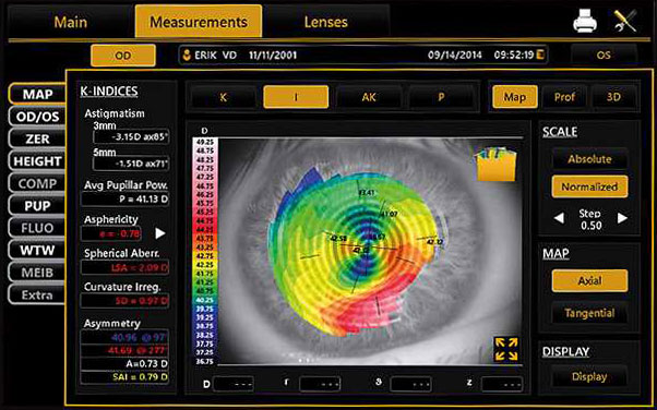
Corneal topography is a painless non-contact computer-assisted diagnostic tool that creates a three-dimensional map of the cornea. The cornea is responsible for the majority of the eye’s focusing power. An eye with normal vision has an evenly rounded cornea. If the cornea is too flat, too steep, or unevenly curved, vision is impaired. The greatest advantage of corneal topography is its ability to detect irregular conditions that cannot be detected by most conventional testing techniques. This type of analysis provides our doctors with very fine details regarding the condition of the corneal surface. These details are used to diagnose, monitor and treat various eye conditions.
Computerized corneal topography can be beneficial in the evaluation of certain diseases and injuries of the cornea including:
- Keratoconus
- Corneal diseases
- Corneal abrasions
- Corneal deformities
- Irregular astigmatism following corneal transplants
It is also an essential tool for specialised contact lens fitting such as Ortho-K and RGP’s (hard lenses). Our doctors will use it to assess the ocular surface prior to contact lens fitting, allowing us to observe how a contact lens alters the shape and quality of your cornea and the tear layer.
Sources:
1 “The Benefits of optomap”, Retrieved 13 July 2019. <https://www.optos.com/en/products/the-benefits-of-optomap/>.
Images courtesy of optomap