EYE EXAMS AND CONTACT LENS FITTINGS
Seeing the Unseen Signs of HealthEye Exams
(Click on tabs to view more information.)
- Children's Eye Exams
- Adult Eye Exams
- Seniors Eye Exams
- AMD Exam
- Cataract Exam
- Diabetes Eye Exam
- Glaucoma Exam
Growing Eyes Can Change Quickly. Annual Eye Exams for Children Are Important- Book Your Child’s Eye Exam Today >

All parts of the body grow as we mature. For children, this process is accelerated as their growing bodies enter adolescence and puberty. Just as they grow in height and weight, so do their eyes: they become longer, wider, and change with maturity. Refractive errors – such as myopia, hyperopia, and astigmatism – are a result of a change in the shape of the eye or its various structures (most notably, the cornea). While someone may not be born with a refractive error, it can absolutely develop over time.
YOUR CHILD CAN’T TELL YOU WHAT THEY DON’T KNOW
A child is born with an adaptable mind. Coupled with a lack of life experience, many children simply aren’t aware that the blurriness or difficulty they face in their vision is abnormal. Therefore, they don’t know how to articulate what they’re seeing. After all, they don’t know anything different.
SETTING YOUR CHILD UP FOR SUCCESS
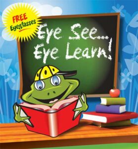
At Listowel Vision Care, our main focus is ensuring your child enjoys a lifetime of great vision. We know how essential vision is to a growing child – especially academically. Remember, the Province of Ontario provides all children under age 19 annual financial coverage for their eye exams. Book your child for their annual eye exam today.> In addition to this, Listowel Vision Care participates in the Eye See…Eye Learn® program which provides complimentary glasses to children headed to kindergarten. Only 16% of children in Ontario have their eyes examined prior to kindergarten. Be sure that your child is one of them! If your child needs eyeglasses, a pair will be provided at no charge (in partnership with Modern Optical Canada and Plastic Plus). We are proud to participate and to provide our community’s young students with the eye care they need to succeed in school. Learn more about the Eye See…Eye Learn Program here >
FACILITATING ACADEMIC SUCCESS
Over 80% of what we learn in school is presented visually – As you can imagine, difficulties with eyesight can have a heavy influence on how your child performs academically. An eye exam is all it takes to ensure your child has the tools they need to succeed.
WHAT TO EXPECT DURING THE EYE EXAM
Be assured that our eye exams are non-invasive and are completely painless. If your child is nervous, let them know that everything we do is safe and gentle. During the exam, we will perform a series of diagnostic tests. These tests look for things such as refractive errors and other conditions that may be impairing their vision or represent a potential health risk. The diagnostic tests typically take between 15 and 20 minutes. With the preliminary tests completed, you and your child will meet with one of our Optometrists. The Optometrist will review the information collected from the tests as well as perform a few of their own. During this time the Optometrist will also assess other areas of health that impact eyesight and provide information and next steps (as needed).
TESTS PERFORMED DURING THE EYE EXAM
- Preliminary Tests – These tests are usually performed by an optometric technician prior to seeing our Optometrist.
- Digital Retinal Optomap – An image of the back of the eye detects signs of diabetes, macular degeneration, optic nerve health, and cataract formation.
- Ocular Coherence Tomography (OCT) imaging – A highly-detailed image of the optic nerve and retina provide essential diagnostic information about eye health.
- Visual Field Analyzer –This test assesses your child’s visual field.
- Auto-Refractor – Reads the curvature of the eye and helps approximate prescription readings. The measurements taken by the auto-refractor guide the Optometrist in fine-tuning the corrective lens prescription (if needed).
- Non-Contact Tonometer – We usually only perform this test on children over the age of ten, unless there is a family history of Glaucoma. This test is performed by a measuring device that monitors the pressure of the eye. Abnormally high intraocular eye pressure (IOP) is a known indicator of glaucoma.
- Slit Lamp Exam – Using an intense light and a specialized magnifying lens, the Optometrist assesses the sclera, cornea, and retina for signs of disease or abnormalities.
- Visual Acuity Test – The famous “letters on the wall” test. The Optometrist will ask your child to read back letters and numbers from a standardized distance (approximately 20 ft / 6 m) in order to determine their visual acuity. For the very young child, we use completely objective acuity measurements, so the doctor does not require a response from the child to assess the sharpness of their sight. In slightly older, but not-yet-reading children, we utilize several different modalities and targets to achieve the sight measurement.
- Retinal Imaging Assessment – The Optometrist will review the images taken by the digital retinal camera and OCT. Although we are not expecting to discover sight-threatening situations in children, a baseline is very important, so that we can determine whether a change occurred, either as a result of an injury, concussion or disease, or even nutritional challenges.
If it has been a while since your last eye exam, schedule yours today. >

Your eyes are incredible organs – perhaps some of the most incredible of all creatures on Earth – and assessing them can tell us more than simply how well you see (though that’s certainly important). As part of our comprehensive eye exam, we also look for signs of other diseases (such as diabetes) that may not be on your radar. Most of our eye exams are completed in under an hour and will detect any changes to your sight and eye health.
What We’re Looking For
Changes to your sight – Most eye diseases that are sight-threatening, such as glaucoma and diabetic retinopathy, are slow to develop. The impact of these diseases on your sight are subtle, often “creeping” over time. By the time you notice something is different, the loss of sight is typically irreversible. By identifying these changes early we are 95% more likely to preserve your visual acuity. Developing eye diseases – Glaucoma, macular degeneration, and other eye diseases have tell-tale signs that can be found via our exams. By utilizing advanced equipment that precisely image your eye (our ocular coherence tomography equipment, used to check for glaucoma, is accurate to within 10 microns), we can detect and treat these diseases in their infancy. Other signs of your overall health – Our patients are surprised to learn that Optometrists often detect diabetes before their family doctor. Why? Because your eyes display tell-tale signs that our Optometrists are specially trained and equipped to detect.
WHAT TO EXPECT DURING YOUR EYE EXAM
Our eye exams are comprehensive, thorough and accurate. For the healthy Canadian adult, an eye exam every one to two years is sufficient. If you have diabetes, Glaucoma, or wear contact lenses, annual eye exams are recommended.
TESTS WE PERFORM
Preliminary tests – These tests are usually performed by an optometric assistant (CCOA) prior to seeing our Optometrist.
Digital Retinal Optomap – An image of the back of your eye detects signs of diabetes, macular degeneration, optic nerve health, and cataract formation, as well as high blood pressure, cholesterol, and other health concerns.
Ocular Coherence Tomography (OCT) imaging – A highly-detailed, 3D image of your optic nerve and macula provide essential diagnostic information about your eye health.
Visual Field Analyzer –This test assesses your visual field and is useful for indicating macular degeneration and glaucoma, in addition to tumours and stroke symptoms.
Auto-Refractor – Reads the curvature of the eye and helps approximate prescription readings. The measurements taken by the auto-refractor guide the Optometrist in fine-tuning your prescription.
Non-Contact Tonometer – A measuring device that monitors the pressure of the eye. Abnormally high intraocular eye pressure (IOP) is a known indicator of glaucoma.
Optometrist-Performed Tests – These tests are performed by the Optometrist. If needed, the Optometrist may perform other tests in addition to the tests below.
Slit Lamp Exam – Using an intense light and a specialized magnifying lens, the Optometrist assesses your sclera, cornea, and retina for signs of disease or abnormalities.
Visual Acuity Test – The famous “letters on the wall” test. The Optometrist will ask you to read back letters and numbers from a standardized distance (approximately 20 ft / 6 m) in order to determine your visual acuity.
Retinal Imaging Assessment – The Optometrist will review the images taken by the OPTOMAP and OCT.
Many Eye Diseases Become More Prominent With Age. Schedule Your Annual Eye Exam Today>

HOW DO OUR EYES CHANGE AS WE AGE?
It seems that all things change over time, and of course, our eyes are no exception. Like the rest of our body, our eyes change in shape, size, and capability. Lenses that were once clear become clouded; cells that were once flexible become more rigid – due to simply advancing years or due to Sun-UV exposure, computer screen exposure or health challenges.
Some changes, such as presbyopia, are natural and unavoidable while others, such as cataracts, can be treated successfully and your visual acuity restored.
PRESBYOPIA & REFRACTIVE ERRORS
Refractive errors are what cause the need for corrective lenses. You may have heard of these as myopia (nearsightedness), hyperopia (farsightedness), astigmatism, and presbyopia. A refractive error is when your eye or part of your eye (usually the cornea) is shaped or sized in a non-ideal manner. This changes how light focuses in your eye, impairing your vision. In some cases, there are distorted parts of the eye that also challenge the ability for sharpness and ease of sight.
INCREASED CHANCE OF VISION – THREATENING EYE DISEASES
Many eye diseases, such as glaucoma and macular degeneration, become more probable as we get older. The prevalence of these diseases is a big part of the reason why the Ontario Association of Optometrists (OAO) strongly advise senior citizens to have their eyes examined every year (as opposed to every other year, as recommended for healthy people between ages 20 and 65).
Many eye diseases don’t have symptoms. Detecting them before they cause sight loss is critical in maintaining your visual acuity and comfort and efficiency of vision.
MORE INFORMATION ABOUT EYE EXAMS
An eye exam is a painless and non-invasive way to keep a pulse on your health. Your eyes are fragile organs that tend to do their job without much in the way of complaints. Checking in every year ensures that we are on top of any changes or diseases that may affect your vision, and often also, other parts of your body.
Most eye exams are completed in under an hour. Schedule your appointment today>
WHAT EYE EXAMS ARE LOOKING FOR:
A change in the health of your eye/internal structures – While everything may look good from the outside, the real action is going on inside the eye. During the eye exam, we use sophisticated retinal imaging equipment that allows us to obtain high-resolution images of your eye and optic nerve.
Developing eye diseases – Glaucoma, macular degeneration, and other eye diseases can take weeks, months, and sometimes even years to develop to a state where the patient notices it. By detecting these diseases before they cause vision loss, we can improve the odds of vision preservation by as much as 95%. Establishing early baseline measurements is crucial to determining change.
A change in your corrective lens prescription – Having to squint more to read? Getting headaches when concentrating? It is a common occurrence that these complaints are remedied by updating your corrective lens prescription. This is taken care of during the exam.
TESTS PERFORMED DURING THE EYE EXAM
Preliminary Tests – These tests are usually performed by an optometric assistant (CCOA) prior to seeing your Optometrist.
Digital Retinal Optomap – An image of the back of your eye detects signs of diabetes, macular degeneration, optic nerve health, and cataract formation.
Ocular Coherence Tomography (OCT) imaging – A highly-detailed 3-D image of your optic nerve and retina provide essential diagnostic information about your eye health.
Visual Field Analyzer –This test assesses your visual field and is useful for indicating macular degeneration, glaucoma and also is important in assessment of concussion or stroke.
Auto-Refractor – Reads the curvature of the eye and helps approximate prescription readings. The measurements taken by the auto-refractor guide the Optometrist in fine-tuning your prescription.
Non-Contact Tonometer – A measuring device that monitors the pressure of the eye. Abnormally high intraocular eye pressure (IOP) is one of the indicators of glaucoma.
Optometrist-performed Tests – These tests are performed by the Optometrist. If needed, the Optometrist may perform other tests in addition to the tests below.
Slit Lamp Exam – Using an intense light and a specialized magnifying lens, the Optometrist assesses your sclera, cornea, and retina for signs of disease or abnormalities.
Visual Acuity Test – The famous “letters on the wall” test. The Optometrist will ask you to read back letters and numbers from a standardized distance (approximately 20 ft / 6 m) in order to determine your visual acuity.
Retinal Imaging Assessment – The Optometrist will review the images taken by the digital retinal camera and OCT.
Age-Related Macular Degeneration Exam
We Are Compassionate Age-Related Macular Degeneration Practitioners Located in Listowel.
HELPING MANAGE YOUR AMD
For millions of Canadians, age-related macular degeneration (AMD) has a very personal impact on their lives. In Canada, macular degeneration is the leading cause of significant vision loss. Working together, we can minimize the impact AMD will have on your vision and help you retain the best level of visual acuity possible.
Learn more about Age-Related Macular Degeneration here >
HOW WE HELP
- We determine the type & severity of your AMD – Macular degeneration has two types: dry (atrophic) macular degeneration and wet (exudative) macular degeneration. During our comprehensive eye exam we will assess your age-related macular degeneration. We also determine the severity of the disease. Staying on top of its development allows us to better predict and manage its effects.
- We help you minimize & manage vision loss – Through corrective lenses, laser surgery, and using other assistive devices, we can help you compensate for the changes to your vision that AMD causes. Our Optometrists and eye care team work to improve our patient’s quality of life in every way we can.
Diagnosing AMD
During an eye exam we will look for signs of AMD. There are numerous tests that we perform that indicate the growth of AMD.
- We use the VitaRisk, a state of the art test that assesses your genetic risk profile. This test can help inform treatments for AMD, as well as determining the probability of the disease progressing to wet AMD.
- The Amsler grid test is a very famous and simple to perform test that involves you looking at a grid of vertical and horizontal lines. In the middle of this grid is a dot; patient’s cover one of their eyes, using the free eye to focus on the dot. The appearance of the grid (such as appearing wavy, blurry, or dark) can indicate the presence of AMD.
- Digital retinal imaging is a process where we take high-resolution images of your inner eye, including your retina and macula. Signs of macular degeneration can be easily spotted and tracked.
- Ocular coherence tomography (OCT) scans and maps your retina. This process produces a detailed image of your macula and provides valuable information about cellular thickness and health.
Cataract Exam and Management
Visit Us in Listowel To Learn if Cataract Removal Surgery is Suitable for You.
Learn more about cataracts here >
WE’LL DETERMINE IF YOU’RE A VIABLE CANDIDATE FOR CATARACT REMOVAL SURGERY
Cataract removal surgery is the only method available for permanent cataract correction. This surgery is relatively painless, minimal hassle, and quick to recover from. However, not all people are candidates for surgery.
ARE YOU A SURGICAL CANDIDATE?
There is only one way to permanently address vision impairment caused by cataracts: remove them. Cataracts are not a “disease” so much as they are the gradual discolouring of the lens in one or both eyes. This means that the eye cannot “heal” and cure the cataract on its own.
Instead, the cataract-stricken lens is removed and replaced with an artificial intraocular lens (IOL).
AN OVERVIEW OF CATARACT REMOVAL SURGERY
Cataract removal surgery is one of the most frequently performed (and safest) procedures available in Canada today. This outpatient procedure is fast, has minimal risk of complications, and provides a permanent solution for cataracts.
Once we have determined your candidacy for surgery we will refer you to a local Ophthalmologist (who will perform the surgery).
Together, we will select a new intraocular lens (IOL) to replace your natural lens. The IOL is used in place of the natural lens and is not seen or felt once inserted. Different types of IOL’s are available for different needs.
When surgery begins, the eye is numbed so that no pain is felt. The surgeon then uses radio waves to break up the natural lens, which is removed via suction and disposed of. The IOL is inserted in its place.
The surgery is quite fast, with the procedure itself taking between 10 and 20 minutes. Recovery is also quite quick, with discomfort largely minimized via eye drops and topical medications.
WHERE WILL YOUR SURGERY TAKE PLACE?
There are several surgeons that our office will refer patients to. Dr. Armogan and his staff at The Ophthalmic Consultant Centre (OCC) in Mississauga, ON is highly recommended for cataract surgery. The OCC are able to schedule consultation appointments promptly, typically within two weeks.
Don’t Let Diabetes Harm Your Vision. Visit Us in Listowel for a Comprehensive Dilated Eye Exam.
DIABETES CAN IMPACT YOUR VISION WITHOUT YOU KNOWING… UNTIL IT’S TOO LATE
For people living with diabetes, preemptive care is the best way to ensure their eyesight remains healthy. As it does with other parts of your body, diabetes takes a toll on your eyes; diligence is required in both planning and care to ensure that your visual acuity remains the best it can be. This is where we can help.
The Ontario Association of Optometrists (OAO) strongly recommends that people living with diabetes have their eyes examined every year (or as frequently as recommended by the Optometrist).
Learn more about Diabetes and the Eyes here >
HOW WE HELP MONITOR & PRESERVE YOUR EYESIGHT
- Our dilated eye exams utilize today’s most advanced diagnostic and imaging equipment. Using it, we take high-resolution images of your eye and monitor any change in its health.
- We have considerable experience and expertise working with patients that have diabetes. We leverage this experience to benefit our patients as much as possible.
- Proactive eyecare allows us to detect any changes to your eye health before they cause vision loss. As vision lost cannot be recovered, preemptively detecting and treating these changes ensures the best possible chance for long-term vision quality.
HOW DIABETES INFLUENCES YOUR VISION
If you have diabetes and have not had an eye exam in more than a year, please schedule an appointment and meet with one of our Optometrists. Your eye health is important in preserving your vision- see us today!
Explaining ‘Diabetic Eye Disease’ – The term ‘diabetic eye disease’ is used to describe a series of eye diseases that are negatively influenced by diabetes. These diseases include glaucoma, cataracts, and diabetic retinopathy. When most people say “diabetic eye disease”, they are probably referring to diabetic retinopathy.
Diabetes can cause these diseases to develop faster than they would in someone without diabetes. In the case of diabetic retinopathy, only people that have diabetes can develop it. All of these diseases influence vision, though both glaucoma and retinopathy do so in a mechanism that is very different than how cataracts impair your sight.
Diabetic Retinopathy – Of the diseases associated with diabetes, retinopathy is by far the most well known. Diagnosed via a dilated eye exam, retinopathy is a vision-threatening disease that can have serious and irreversible impacts on your eyesight if it is not diagnosed and managed appropriately.
Abnormalities in blood vessels in the retina cause retinopathy. Diabetic retinopathy has two classifications broken down into several stages: non-proliferative and proliferative retinopathy.
- Non-proliferative diabetic retinopathy – Broken down as mild/moderate/severe, non-proliferative retinopathy is marked by the blockage of blood vessels in the retina. As the blood vessels become blocked, cellular starvation occurs in retinal cells.
- Proliferative diabetic retinopathy – The final and most severe stage of retinopathy, proliferative diabetic retinopathy is marked by the formation of new blood vessel growth. During the earlier stages of retinopathy, the blockage of blood vessels causes the body to release compounds that will promote new blood vessel growth. This sounds like a good thing, but due to the relative weakness of these blood vessels, they wind up dying and leaving behind scar tissue. This scar tissue pulls at the retina, potentially causing a retinal detachment.
Symptoms of Diabetic Retinopathy
- Blurry vision
- Areas of vision that are heavily obfuscated, as if looking through droplets of water
- Dark spots in one or both eyes (sign of retinal bleeding)
- Blind spots in your normal field of vision
- Difficulty reading/focusing due to impaired central vision
Diabetic Macular Edema (DME) – Only people that have diabetic retinopathy can develop diabetic macular edema. Fluid that leaks from damaged blood vessels (due to retinopathy) will eventually cause retinal swelling- this is DME.
Symptoms of Diabetic Macular Edema
- Distorted central vision (as if objects are wavy)
- Decrease colour differentiation (colours appear faded and washed out)
We’re Here to Help Diagnose & Manage Your Glaucoma. Serving Listowel Since 1949.
ACCURATE DIAGNOSES & ADVANCED TREATMENTS FOR GLAUCOMA
We are proud of our legacy as one of Listowel’s leading eye care clinics. Since 1949 we have focused on one thing: offering our patients the best care possible. By incorporating new equipment and modern diagnostic techniques, we are able to detect glaucoma weeks and months before what was possible even just a few short years ago.
Learn more about Glaucoma here >
GLAUCOMA MANAGEMENT STARTS WITH A CONVERSATION
If you have glaucoma, or are worried that you may have glaucoma, please do not wait any longer for an assessment. Vision that is lost to glaucoma cannot be restored; prevention and preservation is our sole focus. Request an appointment to have an Optometrist assess your glaucoma.
Our management for glaucoma centres along three main points:
- Accurate diagnosis & monitoring – Before we can manage glaucoma, we need to understand the type and severity of it. We will examine the eye regularly and look for changes in eye health; in particular, we will assess the health of the optic nerve.
- Management therapy – There are numerous ways glaucoma can be managed. Before we get into anything surgical we use medicated eye drops and oral medications. These treatments centre around reducing your intraocular pressure (IOP), and thus minimizing potential damage to the optic nerve.
- Advanced management – If your case of glaucoma is proving difficult to manage conventionally, we may consider employing surgical methods that can help reduce intraocular pressure. The most common surgical procedure for managing glaucoma is a laser trabeculoplasty, where a laser is used to carve a small hole near the drainage angle of the eye. This allows eye fluids to drain as normal, resulting in lower IOP.